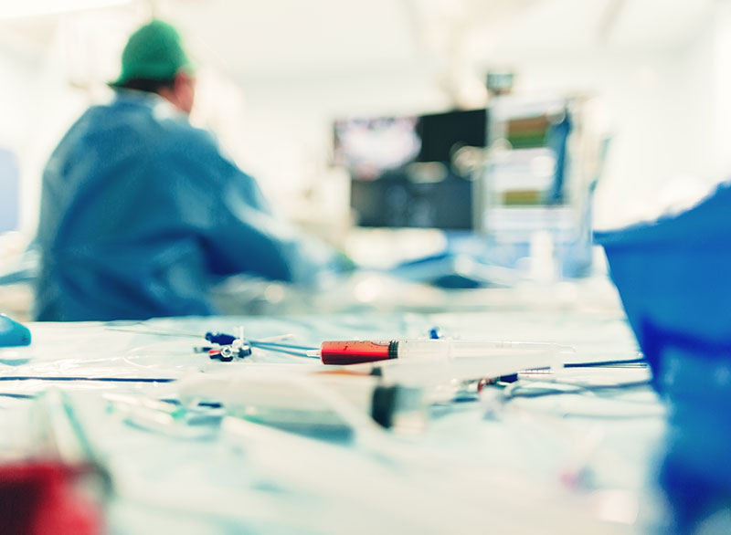
Without the need for surgery or major operation from almost any tissue or organ in the body...
Stenosis and occlusion of the arteries
Artery occlusion usually occurs due to a disease called vascular calcification or atherosclerosis.
It can be seen in anyone, but it is more common in elderly patients, diabetics, people with high cholesterol and blood pressure, and smokers. Arterial occlusions create problems in the region of the organ involved. When using the arm in the arm vein, there are complaints such as fatigue, high blood pressure in renal vascular stenosis, clotting in the brain in carotid artery stenosis. It is possible to treat almost all vascular stenosis and occlusion with angiography.
Leg vein occlusions
Leg vein occlusion is a very common condition, especially in older people. Like other vascular occlusions, it often develops due to atherosclerosis. In a patient with leg vascular occlusion, walking in the early period causes pain in the calf muscles. If the disease (occlusion) progresses, it can cause coldness and severe pain in the fingertips, finger wounds and gangrene.
If the following symptoms are present, there is a high probability of vascular occlusion in the legs:
- - Pain in the calf muscles below the knee with walking
- - The pain is sometimes felt as cramping, sometimes as a stiffening of the muscles
- - Pain does not occur while sitting or standing, it is only with walking
- - The pain that occurs when walking is completely relieved by resting for 2-3 minutes
- - When you walk again, there is usually pain again at the same distance
If you have these complaints, you should be evaluated in terms of leg vein occlusion. Apart from the examination, the first evaluation is usually done by color Doppler ultrasound. If necessary, angiography with tomography or MR is performed (simple angiography).
Today, the majority of vascular stenosis and occlusion can be successfully treated with angiography. Many vascular occlusions, which are said to be incurable, can actually be treated with angiography. It's usually about experience and making good use of existing technology. Our center is one of the most experienced centers in the country in this regard. In our center, all methods such as balloon, medicated balloon, stent, medicated stent, thrombectomy (clot removal) and atherectomy (vein shaving) are used for the opening of arterial stenosis and occlusion. For more detailed information: You can refer to www.leventoguzkurt.com.
Diabetic foot wound
If non-healing wounds occur in the foot in diabetic patients, approximately half of these are due to leg vein occlusion. Foot wounds usually start with simple causes such as bumps on the toes and heel, cutting nails, hitting shoes. Wounds may not heal for weeks or months. Especially in wounds that do not heal for a long time, it should be evaluated whether there is a blockage in the leg veins. If foot wounds are not treated in time, it can cause loss of the leg (amputation).
In a patient with a diabetic foot wound, arterial occlusions are neglected and wound treatment is applied without opening the occlusion. Vascular occlusion disrupts the nutrition of the leg, prevents the wounds from getting nutrients and oxygen, and the wound healing time is prolonged. For this reason, the leg arteries of every patient with a diabetic foot wound should be evaluated and if there is an obstruction, these blockages should be opened first. For treatment, angiography and vascular opening methods are preferred because the risk is low and the chance of success is high. Our center is highly experienced in this regard.
Buerger's Disease
It is an occlusive disease of the arteries that develops especially in male patients aged 20-40 due to smoking. It mostly affects the leg vein, sometimes the hand vein. Usually, due to leg vascular occlusion, there is pain in the leg muscles or sole of the feet and prevents walking. In advanced stages, there are severe pain and wounds in the feet and hands, especially at rest. In a significant part of the patients, it may be necessary to amputate the toes or feet due to non-healing wounds and gangrene. There is no definite cure. The most important element in treatment is smoking cessation. If smoking is stopped, the progression of the disease usually stops. However, the veins that have been clogged up to that point will never open spontaneously. There is no drug treatment either. Very few patients are suitable for surgery (bypass surgery).
The treatment method with angiography is unknown to patients and even most doctors. For this reason, patients are told that there is no chance of treatment and they only need to use drugs. However, in these patients, angiography is the most effective treatment method in opening the clogged vessels. However, Buerger's disease is different from other vascular occlusions. Generally, there is occlusion in the small veins below the knee and it is more difficult to open these veins with angiography compared to other vascular occlusions. It may not be possible to open the veins in every patient. prof. Dr. Levent Oğuzkurt is one of the first and most experienced doctors in our country to treat Buerger's disease with angiography.
Carotid artery stenosis of the neck
In the neck arteryAki strictures can cause blood clots to form in the brain and cause a stroke. If the narrowing is mild, that is, if there is less than 50 percent narrowing in the vessel, the risk is low and drug treatment is sufficient. If the narrowing of the neck artery is more than 50 percent, the probability of clotting in the brain is higher and the stenosis usually needs to be corrected or removed.
Which methods are used to open vascular occlusion?
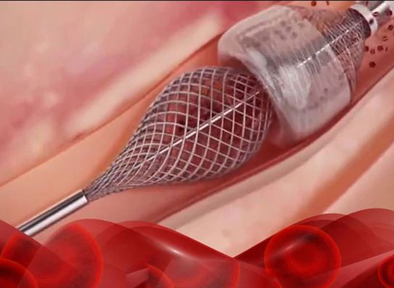
1. Balloons and medicated balloons: These are the vascular opening methods used during angiography. During angiography, a thin and long balloon is placed in the blocked or narrowed vessel, the balloon is inflated to the diameter of that vessel and the stenosis is opened. There is a balloon size for each vein. The balloon is deflated and taken out. So the balloon does not stay in the body.
Medicated balloons are the outside of normal balloons coated with a special drug. When the balloon is inflated, this drug passes to the vessel wall and reduces the possibility of re-narrowing or occlusion of the vessel in the long run.
2. Stent and medicated stent: Stents are different from balloons. They are round metal braided structures. It is like a pipe but not completely closed. Stents are also placed in the narrowed or occluded vessel during angiography and open the vessel. Unlike balloons, stents remain in the vein and cannot be removed.
Medicated stents are just like normal stents. A special drug is placed on the stent, as in medicated balloons. This medicine reduces the chance of re-narrowing or blockage of the vessel in the long term. These are also currently only available for leg veins.
3. Vascular shaving (atherectomy) method: Vascular shaving is to clean the occlusive tissue in the narrowed or occluded vein by shaving with a special device. The device takes the shaved tissue into its chamber and the tissue is thrown out. Vascular shaving can be used alone, or a balloon, medicated balloon or stent can be applied to the corrected vein after shaving. The aim is to keep the vessel open in the long term.
Aortic aneurysm (chest/abdominal main artery ballooning)
Aneurysm is the enlargement or ballooning of the vessel. An aneurysm can occur in any vessel of the body. An aortic aneurysm is the ballooning of the largest artery in the body (in the chest and abdomen). Aortic ballooning usually does not cause any complaints and is discovered incidentally. If left untreated, they can rupture, causing severe internal bleeding and often death. In our center, aneurysm treatment can be successfully performed by placing a stented artificial vessel (stentgraft) into the enlarged vessel by angiographic methods without open surgery.
Myoma treatment (Myoma embolization)
Myoma is a benign tumor that develops in the uterus. According to statistics, fibroids can be seen in one out of every three women over the age of 35. There may be one or more fibroids in the uterus. As fibroids increase in size and number, they cause menstrual irregularity, prolongation of menses, anemia and painful menstruation.
While surgical removal of the entire uterus is often performed in advanced ages, uterine fibroids can be treated both surgically and non-operatively by embolization in the younger age group. In this technique, this vessel is blocked by entering through the artery feeding the fibroid. Myoma begins to shrink because it cannot get enough nutrition. There is no problem in uterine nutrition. The most important advantage of the treatment over surgery is that there is no surgical incision, it does not require general anesthesia, the procedure can be repeated in case of recurrence of fibroids, and the uterine tissue is preserved.
Varicocele treatment
Varicocele is varicose veins that drain the blood in the testicles by enlarging for various reasons. Varicocele is mostly in the left testis (about 85 percent) and bilateral in the remaining patients. It may not cause any problems. It can cause pain in a small group of patients. More often, it can reduce sperm count and lead to infertility. It is mostly treated for this reason. There are two treatments: Surgical closure of these vessels or occlusion of this vessel with angiography (which is simpler, vein angiography).
Varicose veins and modern treatment methods
Varicose veins is an extremely common vein disease. It is more common in women. All types of varicose veins can be successfully treated with new methods such as laser, radiofrequency (RF), steam and foam developed in recent years.
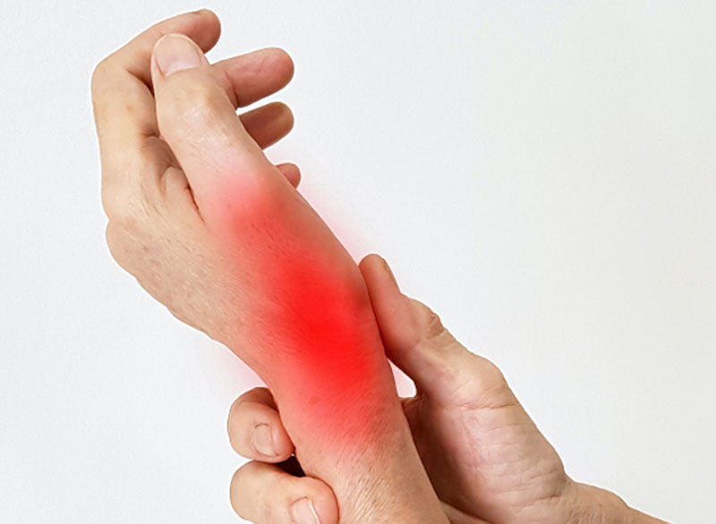
Vein occlusion (Deep Vein Thrombosis or DVT) and its treatment
Vein occlusion can occur in any vein, but the most common and most important one is in the leg vein. If the leg vein is blocked by coagulation, it becomes difficult for the blood to return to the heart.accumulates in the legs and causes swelling of the feet, sometimes causing pain. Mostly in one leg, but can involve both legs. The more vessels the disease takes, the bigger the problem.
It is very important to diagnose the disease early and treat it well in the early period. Because if it is not treated well in the early period, it can create lifelong problems that do not improve and have no treatment.
DVT can be seen in everyone, but the following features increase its incidence: Genetic tendency to clot, having a deep vein thrombosis before, some types of cancer, advanced age, pregnancy and post-pregnancy, use of birth control pills, major complications such as knee and hip prostheses. after the surgery, after the foot is placed in a cast in case of foot fractures and soft tissue injuries.
There are two stages of DVT disease:
1. Early stage (acute) deep vein thrombosis: In the early stage, the clot is fresh. There is foot swelling due to occlusion of the vein and is evident. In one of three patients, the clot in the leg can go to the lungs. This poses a life-threatening risk. In the treatment, blood thinners are used, but these drugs do not clear the clot, they can only prevent new clots from forming and throwing the clot into the lungs. The only treatment that can clear the clot is intravenous therapy in the angiography room. If the clot has reached the inguinal region, intravascular intervention with angiography becomes necessary. Treatment should be done as early as possible. Because the perfect treatment with the intravenous method is only possible when the clot is fresh. In the first 15 days of the disease, it is possible to completely clear the clot. Then the chance of cure decreases. Angiography, that is, intravenous treatment, is mostly done by entering the vein behind the knee and sometimes the ankle vein. From here, either special clot-dissolving drugs are given with thin plastic tubes called catheters (thrombolysis = cleaning the clot by dissolving) or the clot is mechanically cleaned with special devices (thrombectomy = dissolving the clot by breaking up or taking it out).
2. Late stage (chronic) deep vein thrombosis: A second problem called chronic venous insufficiency develops in the early stage, that is, in about half of the patients who are not treated perfectly in the first month. The clot that cannot be treated in the early period adheres to the vessel wall; it both creates a blockage in the vein and disrupts the vein valves. Both cause lifelong foot swelling. Foot ulcers may develop in some of the patients. It is generally said that there is no chance of treatment and that the patient must live with compression stockings. However, in some of these patients, it is possible to relieve the foot by opening the blockage with the angiography method. Unfortunately, this treatment is hardly known in our country, but our center is very experienced in this regard. In late-stage deep vein thrombosis, clot clearance cannot be performed because the clot has aged and adhered to the wall. Only occluded vessels are opened with balloon and stent. In our center, both early and late-term angiography and intravenous treatments of deep vein thrombosis are successfully applied.
Vascular malformation treatment
It is a vein disease that can develop in every part or organ of the body. It can be called a vein ball or malformation. It is similar but different. It is usually present at very young ages and tends to grow with age. It both disturbs the patient as an image and can cause pain.
There are two treatment methods: surgical removal and needle therapy (ablation therapy). Sometimes both treatment methods are used together. Our department performs needle therapy as a non-surgical method in the treatment of vascular malformations.
Interventional radiology in vascular access problems of dialysis patients
Dialysis fistula vessels may narrow, coagulate and become occluded from time to time, causing arm swelling, numbness or scarring in the fingers. Most importantly, the fistula does not work adequately and the patient cannot enter dialysis. In order to enter dialysis, an intravenous catheter must be inserted, which creates new vascular occlusions in the long term.
What kind of treatment does dialysis patients do in interventional radiology?
1. All narrowing or blockages in fistulas in dialysis patients are treated with angiography. If the fistula is clogged with a clot, the clot is removed by non-surgical clot removal methods. Likewise, if an artificial vessel (graft) is inserted and occluded to enter dialysis, this occlusion is also opened by angiography.
2. Severe arm swelling may occur after opening the fistula in patients who have previously had a catheter inserted in the neck region. This shoulder area is due to occlusion of the vein. The vascular occlusion is removed by opening and arm swelling is corrected.
3. In addition, in hemodialysis patients, if it is necessary to enter dialysis from the catheter in emergency situations, vascular access catheters are placed from each region. Since all procedures are performed under ultrasound guidance, it is safer and less troublesome for the patient.